Shark-Skin Inspired Lattice Structure for Drag Reduction

Sharks are extremely diverse group of vertebrates and inhabit a wide variety of aquatic habitats. The skin of sharks is covered in thousands of tooth-like denticles or scales that are anchored to the collagenous layer of the skin known as the  stratum  laxum.  These  scales  play  an  important  role  in  locomotion in  terms  of drag  reduction  and  lift  production. Despite  numerous  studies  on  the  functional  significance  of  shark  denticles , no  studies  have  been performed  to  investigate  the  effect  of  the  anchoring  of the  denticles  in  the  skin or the effect of denticle shape change on the drag reducing and lifting abilities. Moreover, no studies to date have been conducted to study the effect of the denticles stacking and overlapping on the denticles performance.

The objective of this project is to take  the first steps in understanding the effect of shark denticle morphology(roughness, waviness, texture), stacking, and anchoring on the drag reducing and lift increasing properties of the shark skin using computational models and real-life biomimetic shark skin prototypes.

A compatible literature study is available for this project which is “Review of denticles in sea animals”. The objective of this study is to make an overview of the existing denticles in sea animals, the differences in their design and their influence on the drag reduction for these sea animals.

Interested? Contact Aimée Sakes: A.Sakes@tudelft.nl or Jovana Jovanova: J.Jovanova@tudelft.nl or Mostafa Atalla: M.A.A.Atalla@tudelft.nl

SIGMA Catheter – steering inside the Heart

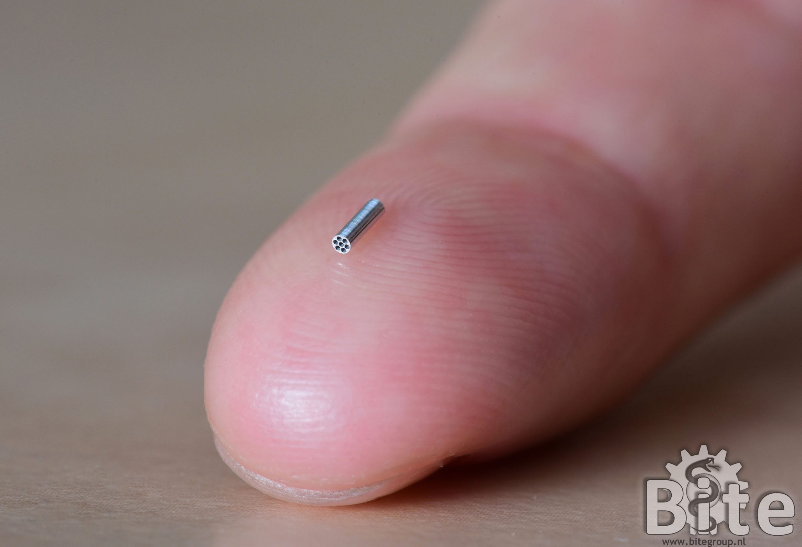
Developed in 2016-2017, diameter Ø3 mm, lumen Ø1 mm

In recent years, steerable catheters have been developed to combat the effects of the dynamic cardiac environment. However, current solutions are bound to a number of limitations: (1) low torsion, (2) shaft shortening, (3) high unpredictable friction, and (4) coupled tip-shaft movements. These effects make it very hard to steer in tortuous blood vessel and inside the heart.

In order to tackle these limitations we developed a novel multi-steerable catheter prototype with four degrees of freedom. The tip has two steering segments that can be steered in all directions, controlled by two joysticks on the handle: one for the thumb and one for the index finger. The prototype features automatic lock of the steering angle once the joystick is released.

To solve the four limitations mentioned above we used eight miniature Bowden-cables inside of the flexible shaft for independent omnidirectional steering of each tip segment. As each segment can steer in all directions, twisting the shaft is not needy for directing the catheter tip, which solves the issue with low torsion (1). The issue with shaft shortening (2) is solved with the Bowden-cables which are axially incompressible. The Bowden cables generate very low predictable friction (3) and coupled tip-shaft movements (4) are absent as the Bowden-cables transfer the joystick motions directly to the tip without influencing the shaft.

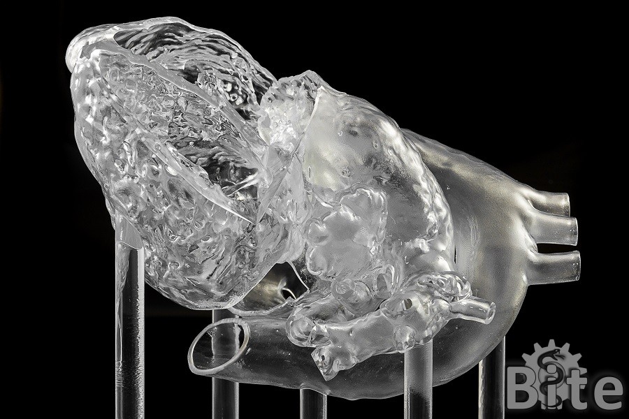
The ability to steer inside the heart with a variety of complex shapes and curves opens great possibilities for complex catheter interventions. We evaluated our SIGMA catheter in a transparent 3D printed heart, based on MRI-images and created by the company Materialize, as well as ex-vivo in a beating porcine heart at the LifeTec Group. Both evaluations show very promising results and superior behaviour as compared to conventional steerable catheters.

Publications:

Ali A., Sakes A., Arkenbout E.A., Henselmans P., Starkenburg R. van, Szili-Torok T., Breedveld P. (2019). Catheter steering in interventional cardiology: mechanical analysis and novel solution. Proc. Inst. Mech. Eng. Part H: Journal of Engineering in Medicine, 12 p.

Media

https://www.materialise.com/en/blog/how-a-modular-testbed-helps-medical-device-developers

Design of a Bone Marrow Harvesting Device (CLOSED)

Bone marrow is a soft and fatty tissue located within the porous bone structure at the centre of the larger bones. During bone marrow biopsies, also called trephine biopsies, a 1-2 cm long sample of the bone and bone marrow is taken to check, amongst others, for blood cell abnormalities. A Jamshidi needle is used to collect the bone marrow by pushing the needle through the rigid cortical outer layer of the bone such that it will be located within the softer cancellous bone. During retrieval of the needle, the biopsy sample sometimes remains inside the incision. This requires manual removal of the sample using forceps, and possibly a second biopsy has to be taken.

During this project, you would first look into current instruments used to perform a bone marrow biopsy after which you will design a novel device that could be used for harvesting bone marrow samples.

Interested? Contact Esther de Kater, e.p.dekater@tudelft.nl

Ovipositor Needle II – Self-Propelling & Steering through Tissue

Developed in 2016-2019, diameters ranging from 1.2 mm to 0.4 mm. 

A wasp ovipositor is a needle-like structure composed out of three elements, called valves. A female wasp uses this structure to drill into wood or fruit and deposit eggs inside a living host. The propagation of the ovipositor through the substrate is achieved by a smart push-pull mechanism, in which one of the valves is pushed while the other two are pulled, using the surface-dependent friction properties with the soft substrate to move forward. Inspired by the ovipositor of parasitoid wasps, we developed a series of self-propelled steerable Ovipositor Needles with ultrathin diameters ranging from 1.2 mm to 0.4 mm.

Our first Ovipositor Needle prototype consists of six super elastic Nickel Titanium (NiTi) wires concentrically arranged around a seventh NiTi wire. The seven wires are interconnected at the tip with a small flower-shaped ring (Ø 1.2 mm) manufactured for minimal resistance during propulsion. The ring has a central hole to which the central wire is glued, surrounded by six concentric holes through which the six other wires can slide back and forth. Each proximal end of the six movable wires is connected to a miniature stepper motor, in which a leadscrew-slider mechanism converts rotational motion into linear motion.

We performed a series of experiments in which the needle was inserted in tissue-mimicking gel phantoms. The wires were sequentially moved back and forth, resulting in the needle moving forward inside the phantom using the surface-dependent friction properties between the wires and the gel. Different sequences of wire actuation were used to achieve both straight, curved and S-shaped trajectories.

In our second Ovipositor Needle prototype we changed the shape of the interlocking ring from cylindrical to conical to investigate the effect of pre-curved wires. We found out that pre-curved wires facilitate steering, however, at the drawback of a slightly larger tip diameter due to the use of a conical flower-ring.

In a final series of Ovipositor Needle prototypes, the flower-shaped ring was replaced by an thin-walled shrinking tube, glued to one of the outer wires, ultimately resulting in ultrathin 0.4 mm needle diameters three times the size of a human hair. The prototypes were tested in multi-layered gel phantoms with varying stiffness properties and artificial membranes, representing different organs and tissues. In a final series of ex-vivo experiments the needles were evaluated with success in porcine liver, kidney and brain tissue.

This project, in which we developed world’s thinnest self-propelled-steerable needles, shows the strength of a novel bio-inspired approach leading to a new generation of needles that can be used to reach deep targets inside the body without a risk of buckling and with the possibility to correct the trajectory. Our needles were developed within the WASP project that focused on the development of steerable needles for localized therapeutic drug delivery or tissue sample removal (biopsy). In a follow-up project, funded by the Netherlands Organization for Scientific Research (NWO) we will develop the needles further towards clinical application in urological interventions under MRI.

(Picture at the top adapted from “Braconid Wasp Ovipositing” by Katja Schulz is licensed under CC BY 2.0.)

Publications

Media

Wasp Inspired Steerable Bone Drill (CLOSED)

Parasitoid wasps can drill through relatively hard material such as wood with their ovipositor, a very thin structure through which the wasp transports its eggs. The ovipositor consists of three valves that slide one-by-one deeper in the wood to drill. Not only can the parasitoid wasp drill with this very thin structure through hard material but the wasp is also able to steer during drilling. It is still not fully known how the wasp can do this, but there are multiple hypotheses. Multiple steerable needles were designed based on the wasp ovipositor. The goal of this graduation project is to design a steerable wasp inspired bone drill.

Interested? Contact Esther de Kater: E.P.deKater@tudelft.nl

HelicoFlex – advancing steering with 3D printing and minimal assembly

Developed in 2019-2020, diameter Ø8 mm

In minimally invasive surgery, instrument maneuverability is limited by the use of small incisions. Increasing the number of degrees of freedom (DOF) of the instrument shaft is beneficial for many surgical interventions. However, increasing DOF usually leads to high mechanical complexity, issues with sterilisation and too large cost price for disposable use.

In an attempt to reduce manufacturing time we propose the first fully 3D-printed handheld, multi-steerable instrument: the HelicoFlex. The instrument is mechanically actuated and is fitted with a compliant shaft containing five serially-controlled segments enabling high maneuverability in 10 degrees of freedom.

Our new, compliant segment design merges the functions of four helicoids and a continuum backbone combining high torsion and axial stiffness with low bending stiffness. Five such compliant segments were combined to form the shaft of the HelicoFlex. Following the control design strategy of our older MultiFlex and HelixFlex devices, a compliant control handle was designed that mimics the shaft structure.

The entire frame of the HelicoFlex consists of only three complex-shaped 3D printed components that are printed without a need for any support material in the compliant section. The use of minimal-assembly 3D printing drastically decreases assembly time. Our 3D printed shaft features four working channels that facilitate combined use with flexible instruments such as biopsy forcipes. With its 10 degrees of freedom, our HelicoFlex showed a fluid motion in performing single and multi-curved paths.

Video adapted from Culmone, C., et al. (2020). Plos one, 15(5), e0232952 licensed under CC BY 4.0

Publications

Entry point detection for spinal fusion surgery (CLOSED)

Multiple diseases can require patients to undergo spine surgery. At the BITE group, we are developing a novel probe that allows for the surgeon to steer through the bone along a secure drilling trajectory, avoiding nerves and blood vessels that run along the spinal column. To help the surgeon find and maintain the right trajectory, an optical sensing system based on Diffuse Reflectance Spectroscopy (DRS) will be integrated into the probe to differentiate the tissue ahead of the tool tip, thereby providing positional feedback for the surgeon in real time.

In the scope of the proposed graduation project, a probe prototype will be designed that enables the surgeon to sense the correct entry point for spine drilling procedures. Its usability for guidance will be assessed through drilling tests on a bone phantom/ex-vivo animal bone.

This assignment will be available from September 2021. Interested? Contact Esther de Kater, e.p.dekater@tudelft.nl or Merle Losch, m.s.losch@tudelft.nl.

Design of an Ultrasound-Enhanced Needle (Closed)

Needles are an integral part of many medical procedures nowadays. In prostate cancer ablation procedures, for example, surgeons insert a needle into the prostate tumor with image guidance to deliver the treatment fiber to the cancerous tissue. Needle insertion is not, however, a simple task and requires precision localization to reach the target accurately. Furthermore, minimal insertion forces are required to preserve the tissue that the needle is penetrating. In nature, some wasp and mosquito species are able to move a needle-like structure in substrates using a vibrating motion, this vibrating motion is thought to reduce the frictional force during the penetration process. In this graduation project, we look forward to developing an ultrasound-enhanced needle to minimize the effect of the friction forces acting on the needle, thus increasing the positioning accuracy and minimizing the tissue damage due to penetration.

The assignment is available from October 2021. Interested? Contact Jette Bloemberg: j.bloemberg@tudelft.nl or Mostafa Atalla: m.a.a.atalla@tudelft.nl.

MemoFlex 1 – Mechanical Surgical Snake

Developed in 2016-2017, diameter Ø5 mm

During complex surgical procedures such as in skull-base surgery, there is a need to reach difficult-to-reach locations via narrow anatomic corridors. Performing surgery along complex 3D pathways requires a snake-like instrument that memorizes the 3D shape of the followed pathway and shifts the shape backward as the instrument moves forward. This snake-like method of locomotion is called “follow-the-leader locomotion”, in which the head is the “leader” and the body follows the pathway of the head, see the following animations:

Follow-the-leader locomotion requires a segmented multi-steerable instrument as well as a memory in which the angles of the segments can be stored and shifted. In robotic approaches, the actuation usually occurs by a range of electric motors controlled by a computer. Although feasible, this will result in a very complex system requiring additional safety measures to ensure reliability during surgery.

In a desire to create a simpler system, we explored an alternative follow-the-leader approach by using a mechanical memory. Following the design approach of our MultiFlex, the MemoFlex 1 contains a 12 cm long, Ø5 mm multi-steerable tip with 14 segments that can be controlled individually in 28 Degrees of Freedom. Using 56 steering cables, the tip is connected to a bendable handle. When the handle is bent in a certain shape, the shape is mirrored and replicated by the tip.

The shape memory is a pre-bent stainless steel rod that slides through the bendable handle, driven by a crank. As the rod slides through the handle, its shape is detected by a 3D-printed compliant helicoid insert that makes the handle follow the shape of the rod precisely. The mechanism replicates the handle-shape to the tip which will then maneuver along a curved pathway equivalent to the shape of the pre-bent rod. The shape of the pre-bent rod can be derived from CT or MRI-images.

Our novel copy-and-replication mechanism shows promising results. Yet, the prototype has a high mechanical complexity. We therefore continued this research with an improved prototype, the MemoFlex 2, which contains aan improved shape memory mechanism and a strongly simplified compliant 3D-printed tip.

Publications

Solving medical problems through nature’s ingenuity