Vacu-SL

In order to fully benefit from the functionalities of flexible endoscopes in surgery, a simple shaft-guide that can be used to support the flexible endoscope shaft is required. Such a shaft-guide must be flexible during insertion into the human body and rigidified when properly positioned to support the flexible endoscope shaft. A shaft-guide called ‘Vacu-SL’ was designed.

The Vacu-SL rigidity control mechanism utilizes the flexural rigidity increase that is achieved by vacuuming foil tubes filled with small particles. In this prototype the influence of particle hardness, size, and shape on the flexural rigidity of vacuumed foil tubes filled with these particles is investigated. Experiments show that the flexural rigidity increases with the hardness and irregularity of the particles and that there may be an optimal particle size in the low particle diameter region.

MicroFlex – Steering at a Micro-Scale

Developed in 2005-2006, diameter 1.3 mm, steering range: ±90º in all directions.

The MicroFlex is a steerable instrument for micro-surgery with a miniature Cable-Ring mechanism consisting of a ring of six steel cables (Ø0.2 mm) surrounded by a spring. The six cables are used for steering the tip, whereas the inner spring is replaced by a central cable that can be used to drive a miniature gripper on the tip (not yet incorporated in this prototype). The result is an instrument that realizes 3D-steering with only seven cables and a spring – smaller and at the same time simpler than existing steerable instruments.


Publications

Media

Endo-Periscope III – Revolution from a Squid

Developed in 2003-2004, diameter 5 mm, steering range: ±110º in all directions.

Construction equivalent to Endo-Periscope II, however with Ring-Springs replaced by novel patented “Cable-Ring” mechanism based on tentacles of squid. The Cable-Ring mechanism consists of a ring of 22 steel cables (Ø0.45 mm) enclosed by two conventional coil springs, allowing only axial cable displacements to control the motion of the steerable tip. The Cable-Ring mechanism is entirely constructed out of standard parts and therefore very suitable for low cost mass production. The Cable-Ring mechanism is being commercialized worldwide by spin-off company DEAM.


Publications

Media

Endo-Periscope II – Smart Ring-Spring Steering

Developed in 2001-2002, diameter 12 mm, steering range: ±125º in all directions.

The Endo-Periscope II has been developed in cooperation with Prof. Shigeo Hirose of the Hirose & Fukushima Laboratory, Tokyo Institute of Technology.

Endo-Periscope II is a simplified, patented version of Endo-Periscope I containing two enhanced compliant Ring Springs to control both left/right and up/down tip rotations. Endo-Periscope II contains an improved spatial parallelogram-mechanism that minimizes bending radius in all positions of the tip. A prominent part of the mechanism is a compression spring in the handgrip that compensates the spring force of the two Ring-Springs.


Publications

Media

Endo-Periscope I – Compliant yet Torsion Stiff

Developed in 1999-2000, diameter 15 mm, steering range: up/down 0º-180º, left/right ±60º

The Endo-Periscope I has been developed in cooperation with Prof. Shigeo Hirose of the Hirose & Fukushima Laboratory, Tokyo Institute of Technology.

Endo-Periscope I is a patented steerable endoscope for laparoscopic surgery containing a novel, torsion-stiff “Ring-Spring”. The compliant Ring-Spring consists of a number of spring-metal rings that are bent and welded to each other in pairs. The rings contain holes for guiding steering cables. The Ring-Spring is controlled by a novel spatial parallelogram-mechanism that unfolds the spring from a compressed position to minimize bending radius of the steerable tip. The tip has always the same orientation as the hand grip, offering intuitive control to the surgeon showing how the tip is oriented in the abdominal cavity. The Ring-Spring is only used to control up/down tip rotations. Left/right tip rotations are controlled by a conventional hinge-mechanism.


Publications

Media

Steerability – How it all started…

During Minimally Invasive Surgery (MIS) surgical instruments and an endoscopic camera are inserted  through tiny  incisions into the human body. This approach reduces the patient’s recovery time, the size of the scars and the risk of complications. Unfortunately, MIS also has its drawbacks since the intervention becomes more difficult for the surgeon:

  • There is limited visual 3D information (and thus no proper depth perception) of the surgical area.
  • The surgeon’s eye-hand coordination is distorted because the camera looks at the organ from a different angle than the surgeon would normally do.
  • The motion of rigid instruments is strongly limited by the fixed position of the incisions.
  • The surgeon’s ergonomic position deteriorates due to the use of  long and rigid instruments in combination with fixed incision points.

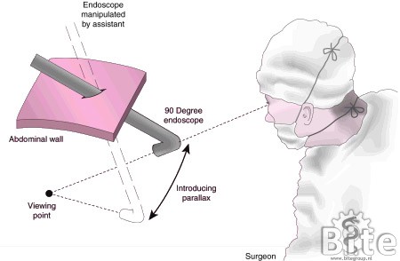
One way to obtain more depth information is to enable motion parallax – obtaining depth information by mutual displacements of objects in the field of view when moving the eye while keeping the viewpoint in focus. With conventional rigid endoscopes, however, moving the endoscope results in the viewpoint to shift away as the endoscope rotates around its incision point.

During his stay in the Hirose & Fukushima Laboratory, Tokyo Institute of Technology, Paul Breedveld and Shigeo Hirose developed an endoscope with a steerable parallelogram mechanism, enabling motion parallax and facilitating eye-hand coordination. This resulted in the first steerable endoscope of the BITE-Group – the Endo-Periscope I.


Publications

Shockwaves for Puncturing through Occlusions (CLOSED)

Chronic Total Occlusions (CTOs) are currently the last frontier for cardiovascular surgery. CTOs are defined as heavily calcified occlusions in the vascular system of the heart that slowly form over time and are at least three months old. The preferred treatment option of CTOs is a so-called Percutaneous Coronary Intervention (PCI). Unfortunately, in many occasions PCI is ineffective as the guidewire is not able to cross the CTO due to lack of stiffness of the guidewire and the less than favourable properties of the CTO. Therefore, the success rate of these kinds of interventions is relatively low, which in turn has led to the tendency to treat these kinds of lesions medically.

To improve the success rate of these kinds of interventions it is, therefore, necessary to develop a new tool that is able to cross the CTO without buckling. From this, the idea arose to use shockwaves to puncture through CTOs. Shockwaves are used in the animal kingdom to strike through hard shells and prey capture by, for example, the Mantis Shrimp. They are particularly effective in fluidic environment such as the blood vessels and are, therefore, a good candidate for prototype development.

This research project will entail a literature study where the use of shockwaves in the animal kingdom and other shockwave applications and/or instruments is investigated, and a graduation project where innovative shockwave tool for puncturing the proximal cap of chronic total occlusions is developed.

Contact: Aimée Sakes, a.sakes@tudelft.nl

Design of A Flexible Tissue Removal Device for Skull Base Surgery

Cushing’s disease is a neurological disorder caused by the loss of dopamine secretion in and near the pituitary gland. Unlike in human Cushing’s disease, where surgical removal of the pituitary tumors is a common treatment modality, in horses oral treatment is the treatment of choice. This oral treatment does not provide a long-term solution to the disease, as it is focused on symptom reduction and does nothing to fight the actual cause of the disease.

In collaboration with the department of Veterinary medicine Utrecht, a new paradigm of surgical treatment of Cushing’s disease in horses has been developed that uses the vascular system in combination with an innovative flexible morcellator to reach the pituitary gland. This flexible morcellator incorporates a rigid tip with a resection tool and a flexible shaft, which incorporates a cable drive element, used for actuating the resection tool, and a central tissue transportation lumen.

Further development of this device is necessary before clinical use can commence. Redevelopment should focus on miniaturisation, improving the cutting blade design, and adding a means of visualizing the pituitary tumors.

We are currently also exploring the possibility to redesign this instrument for human surgery.

Contact: Aimée Sakes, a.sakes@tudelft.nl

Solving medical problems through nature’s ingenuity