Category Archives: Research Projects

NWO – Interventional Ductoscopy – the EVAPORATE study

This research project is funded by the Netherlands Organization for Scientific Research NWO and the Dutch Foundation of Cancer Research KWF.

Ductoscopy is a minimally invasive micro-endoscopic technique that allows for direct visualization of the milk ducts of the breast through their natural orifices in the nipple. It can be performed under local anesthesia in daily outpatient routine and has proven to be safe with a very low risk on (mild) complications. In collaboration with the UMC Utrecht, the aim of this project is to develop novel instruments for ductoscopy to prevent women from getting breast cancer.

TU Delft – Accessible Prosthetics through 3D Printing and a Smartphone App

This research project is part of the Delft Global Initiative program: the portal and booster of Science and Technology for Global Development at TU Delft. Aim of the program is to contribute to sustainable solutions for global societal challenges, through problem-oriented interdisciplinary technical research in close cooperation with partners in the developing world, to meaningfully improve lives of people living in poverty.

NotImpossible-3d-printed-hands-and-arms-1

Combining modern advances in smartphone technology with the seemingly unlimited possibilities of 3D-printing, in this project we aim to create easy access to prosthetics for amputees in Third World countries. We will develop an advanced, free IOS / Android app that scans the amputee with a smartphone camera and completely automates the complex prosthetic design process ending in design drawings for a 3D-printer that manufactures a well-fitting prostheses. In this project we will not only generate new, fundamental knowledge on automatic designing and manufacturing, we will also collaborate with a number of charity organizations to stimulate local initiatives in 3D printing and to optimize the prosthetic supply chain.

Photo: Example of 3D-printed prostheses from Mich Ebelings “Not Impossible Project” in Sudan.

More information: http://globalstories.tudelft.nl/story/paul-breedveld/


NWO – Tree Frog-Inspired Gripping

This research project is funded by the Netherlands Organization for Scientific Research NWO.

Grip between heterogeneous objects can be weak or strong, permanent or reversible, and load dependent or load independent. In some cases, grip needs to be very strong (e.g., aircraft fuselage parts), whereas in other cases it has to be deliberately weak (e.g., screen protection foil for your smartphone). Office tape has to be reversible, whereas a broken vase should be permanently glued back together. A staple can be applied by piercing it through the paper, whereas a surgical gripper should grab soft organs without damaging them.

Grip that is strong, load independent, and reversible at the same time is a great challenge in engineering. Nature, on the other hand, manages quite well: Geckos, for example, can walk on tilted surfaces due to adhesive forces between their toes and the substrate. The stickiness is more than enough to carry the gecko’s weight, but the animal can still easily peel its feet from the surface during walking. Tree frogs can grip on wet, or even flooded, surfaces. Furthermore, their toe pads are soft, thereby eliminating the risk of causing normal stresses to the objects the animal grips upon. With a single foot, tree frogs generate gripping forces strong enough to 100 times carry their weight. Still, tree-frog grip is reversible: The animal detaches from a substrate by peeling its toes off.

The goal of this research project is to gain insight into adhesion on wet and soft surfaces and, inspired by the tree-frog adhesive apparatus, to develop artificial systems that are able to reversibly but firmly grip to objects while minimizing normal stresses.


NWO – Ultra-Thin Steerable Needle for Solid-Organ Interventions – WASP

This research project is part of the iMIT research programme and funded by the Netherlands Organization for Scientific Research (NWO). The iMIT Program, executed by a community of Dutch Universities, university medical centers, and companies, aims to develop instruments for minimally invasive interventions. The program will result in the development of interactive Multi-Interventional Tools (iMIT)  that can adapt to their environment and integrate diagnostic and therapeutic functionalities, thus permitting effective single-procedure interventions.

The WASP project focuses on medical needles – common devices used in minimally invasive percutaneous procedures, such as localized therapeutic drug delivery or tissue sample removal (biopsy). Reaching the target with high accuracy and precision is necessary for the success of these procedures and becomes a challenge when the target is located deep inside the body. The surgeon needs a steerable flexible needle that can follow complex curved trajectories while avoiding sensitive structures, such as blood vessels, located along the trajectory between the insertion point and the target site. Looking in nature we find an interesting behavior in wasps which can be used as source of inspiration for facing this challenge. The wasp has a thin and flexible needle-like structure, called ovipositor, used for laying eggs into larvae hidden inside fruits or wood. It is composed of three longitudinal segments, called valves, that can be actuated individually and independently of each other with musculature located in the abdomen of the insect. In this way the wasp steers the ovipositor along curved trajectories inside different substrates without a need for rotatory motion or axial push.

Inspired by the anatomy and the steering mechanism of this needle-like structure we aim to develop an ultra-thin steerable needle that can follow curved paths through complex solid organs while avoiding obstacles.

NWO – Multi-Steerable Cardiology Instruments – MULTI

This research project is part of the iMIT research programme and funded by the Netherlands Organization for Scientific Research (NWO). The iMIT Program, executed by a community of Dutch Universities, university medical centers, and companies, aims to develop instruments for minimally invasive interventions. The program will result in the development of interactive Multi-Interventional Tools (iMIT)  that can adapt to their environment and integrate diagnostic and therapeutic functionalities, thus permitting effective single-procedure interventions.

Project MULTI – Design and development of multi-steerable tools for cardiac interventions

Cardiac Catheters  
The field of interventional cardiology is a growing branch of cardiology where minimally invasive instrumentation is of high importance. Catheters are among the most versatile and essential instruments used in interventional cardiology. Where in the past they were designed as flexible tubes, meant for monitoring or drug delivery, today catheters have evolved into more complicated and steerable instruments with additional tip functionality. As such, a large variety of commercially available catheters exist, being adopted in treatments of, for instance, heart rhythm defects and heart valve disease.

Current Difficulties
Despite their frequent and essential use, currently existing catheter designs have limited functionality as a result of several difficulties. Precise positioning of the catheter tip in the heart remains one of the biggest challenges as a result of complex 3D shapes inside heart and the absence of vessel wall support. In addition to that, respiration and heartbeat lead to a constant movement of the heart and changes in blood flow inside the cardiovascular system. Therefore, the use of catheters for complex interventions inside the heart requires a catheter tip that can be positioned accurately at the required location without damaging the heart or other surrounding anatomy. Of specific interest are cardiac biopsies and ablations, where mal-positioning of the catheter due to a lack of steerability can results in severe complications such as ventricular perforation or heart block. This research project at the TU Delft therefore focuses on designing and developing a catheter that is multi-steerable and is able to be directed towards and positioned inside any location in the beating heart.

Multi-Steerable Catheter
The aim of this project is to develop new and multi-steerable catheter technology based on the cable-ring technology and human factor experience at the TU Delft. First catheter concepts will allow left/right and forward/backward motion without rotation of the catheter shaft. More advanced concepts will be included with electro-mechanic controls and multiple steering segments that will allow for complex 3D tip motion. Finally, in-vitro evaluation on an isolated beating heart will take place to enable accurate positioning under physiological circumstances. The project is intended to result in explicitly evaluated multi-steerable catheter prototypes that are ready for commercialization. The realization of such a multi-steerable catheter will offer application in more complex minimally invasive cardiac interventions such as annuloplasty procedures, cardiac tissue resections, precision cardiac biopsies, septal defect closures, and valve implantations. Moreover, our focus is on development of steerable catheters for cardiac biopsies and ablations.

NWO – Image Guided Interventional Treatment – IGIT

This research project is part of the iMIT research programme and funded by the Netherlands Organization for Scientific Research (NWO). The iMIT Program, executed by a community of Dutch Universities, university medical centers, and companies, aims to develop instruments for minimally invasive interventions. The program will result in the development of interactive Multi-Interventional Tools (iMIT)  that can adapt to their environment and integrate diagnostic and therapeutic functionalities, thus permitting effective single-procedure interventions.

The IGIT project focuses on increases the success rate of Percutaneous Coronary Interventions (PCI) of Chronic Total Occlusions (CTO). In the field on cardiovascular interventions, the CTO is a subset of lesion types that is the most challenging to treat, evidenced by the low procedural success (55%–80%) depending on the techniques and experience of the physician. In comparison, the procedural success rate of non-occluded lesions is approximately 90%. The most important reason for this lower success rate is the fact that the lesion cannot be crossed by the guidewire. Different techniques with extra equipment and additional guidewires are used to increase the rate of success. E.g., a support catheter can provide extra column strength, supporting the guidewire advancement through the occlusion while maintaining its flexibility. Consequences are an increase of complications such as false lumen creation or vessel dissection. Furthermore, these procedures may take hours leading to high irradiation exposure of patient andthe physician and use of large volumes of nefrotoxic contrast dye.

The aim of the IGIT project is to develop a support catheter with triple functionality: (1) imaging with an ultrasound transducer that does not only look sideways, but also in front, so that the CTO lesion can be visualized and information from the signal can be used to uncover the better accessible areas for crossing the CTO and avoid a dissection of the arterial wall or the creation of a false lumen (ErasmusMC); (2) accurate steering of the device based on the optimal use of the information already given by the location of the device, and the situation in front and around the device (TUDelft); (3) 3D visualization of the catheter without the use of X-ray by integrating a specially designed optical fiber into the support catheter (Philips). Next to these functionalities, several innovative crossing mechanisms that are able to cross heavily calcified CTOs without buckling and with ease will be developed.

In the BITE group we will focus on the design of the steerable catheter. Various methods of steering will be investigated. These include mechanical, electrical, or shape memory materials. A requirement is to leverage the limited space available in the catheter, especially since a part of the space is being used for ultrasound transducers.Furthermore, innovative crossing methods are investigated by looking into nature.

Designs related to IGIT:

  • Pulze Hammer I
  • Pulze Hammer II (coming soon)
  • Accura (coming soon)
  • Volt (coming soon)
  • Wave Catheter (coming soon)
  • Cradle Catheter (coming soon)
  • Biopsy Needle (coming soon)

Publications related to IGIT:

NWO VICI – Dendritic Instruments – Outreaching the Squid

This VICI-research project is funded by the Netherlands Organization for Scientific Research NWO.

Background

In ‘standard’ minimal access surgery, the surgeon inserts rigid instruments through small incisions in the skin or natural orifices to reach targeted areas inside the human body. This approach drastically reduces the invasiveness of surgery compared to conventional open approaches, yet the reduced size of the surgical entry-point does also severely restrict the maneuverability of the used instrumentation. This lack of instrument maneuverability becomes especially apparent when considering Endoscopic Skull Base Surgery (ESBS). A prime target of ESBS are tumors on the pituitary gland positioned at the skull base, the region that separates the brain from the rest of the head. The nose is used as the surgical entry-point, and due to the rigid nature of the used instrumentation, the surgeon needs to create a straight surgical pathway to the pituitary gland. The limited width of this pathway in combination with the need for multiple instruments severely limits sideway movements of the instruments. This situation leads to a phenomenon called swordfighting wherein the shafts of instruments collide and, moreover, it severely restricts the maneuverability of the individual tools (i.e. grasper, scissors, etc.).

We strive to improve on overall instrument maneuverability with the development of dendritic instruments. A dendritic instrument is a maneuverable single-shaft instrument that branches into multiple independently steerable tools. Such an instrument would eliminate the occurrence of swordfighting, as the number or shafts is reduced to one, while providing the surgeon sufficient maneuverability of the individual tools. This research project is divided into two main topics; the mechanical construction and methods of control of dendritic instruments, such that these instruments are able to be implemented in operating theatre in the near future.

Steering

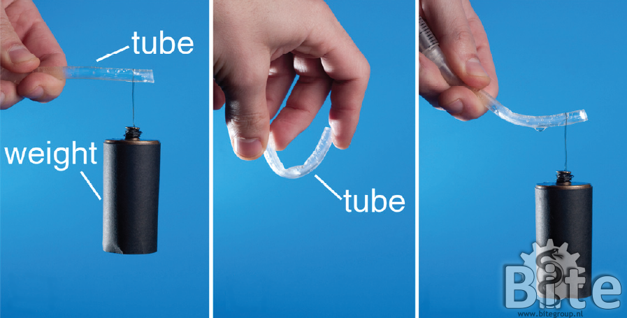
The mechanical construction of a dendritic instrument consists of two basic parts. First, there is the shape memory shaft that should be capable of following a curved trajectory up to the targeted area while providing a stable base. Secondly, there are the individual steerable branches that sprout from this stable base and provide independent maneuverability of each individual tool.

Creating a steerable branch starts with a flexible structure, either containing joints or a compliant backbone. The actuation of such this flexible structure can then be realized by several actuation methods, including the use of electric motors, hydraulic actuators, and shape memory alloys. Our research is focused on a fully mechanical actuation method based on cable-structures. This allows for structures that are easy to miniaturize, eliminates the need for possibly dangerous electric currents, high pressures or high temperatures, and shows high potential for reducing the costs of fabrication.

Besides the obvious requirement that the branches need to be maneuverable, they should also have a certain stiffness in order to cope with external forces that will be present during, for example, tissue manipulation. In our search for a suitable cable-structure to achieve high maneuverability and stiffness, we have developed a cable-structure in which multiple cables are placed at different angles along the longitudinal axis. This structure has already shown great promise and is now in the process of further optimization.

Control

Dendritic instruments consist of many small joints and branches which the surgeon(s) needs to actively steer, in order to perform complex surgical tasks (e.g. suturing or tissue manipulation). The amount of joints in dendritic instruments are even so many, that currently existing prototypes require the cooperation of two surgeons to perform a task which is actually meant for only one surgeon. In other words, dendritic instruments have more Degrees of Freedom (DOF) than any  surgeon can control alone. However, the large amount of DOFs combined with an intuitive method of control is exactly what is required for dendritic instrumentation to become a reality.

The BITE research method to dendritic instrument control is one which is exploration driven. The optimal mechanical construction and DOF configuration are still being researched. Hence, the accompanying control interface or instrument handle cannot be designed yet. To investigate the best methods of control, virtual instruments are simulated in a Virtual Environment (VE). Physical hand movements and gestures are measured with RGB-D Kinect cameras, and mapped to virtual instrument movements. By playing with the coupling between hand DOF and instrument DOF, new control strategies are tested and reverse engineered to ultimately discover the best method for dendritic instrument control.

Dendritic_control_simulation_setup

Image adapted from  www.nimblevr.com (previously www.threegear.com)

Shaft Guidance for Flexible Endoscopes

Flexible endoscopes (long, slender, flexible instruments with a camera and light at the distal end, having working channels to introduce flexible instruments) are used for diagnostic and therapeutic interventions inside the human digestive system and inside the abdomen. Though used for their flexibility, the flexibility of these instruments causes several difficulties during insertion and use. During insertion, flexible endoscopes can buckle and loop, which may hamper full insertion into the patient’s body. During therapeutic interventions, the flexible endoscope fails to provide stability for surgical instruments that are introduced through the flexible endoscope.

Shaft-guidance would be a good solution because it potentially enables following a 3D trajectory without any support of the surrounding anatomy at all. Combining auto-propulsion with a rigidity control mechanism may provide improvement in applications within confined anatomies where auto-propulsion simplifies insertion and rigidifying the endoscope shaft helps to stabilize the instruments during surgery. Three potentially suitable rigidity control concepts are selected and further investigated to quantitatively and qualitatively predict the maximally achievable flexural rigidity of these rigidity control mechanisms:

Vacu-SL

FORGUIDE

PlastoLock

The thesis on this topic can be found on:
Shaft-Guidance for Flexible Endoscopes


FORGUIDE

The FORGUIDE mechanism enables making a shaft-guide out of cheap standard parts that is rigidified by creating a laminate that consists of a spring, cables and expandable tube. The connection between these three layers is obtained by friction. The bench tests showed that the FORGUIDE prototype FGP-01 of only 5.5 mm diameter could provide flexural rigidities up to 1541 Ncm2 , which far exceeds the flexural rigidity of flexible endoscopes. Furthermore, a bending radius of almost 1 cm could be achieved in the compliant state with the FGP-01 without losing the ability to rigidify.

PlastoLock

Flexible endoscopes are used for diagnostic and therapeutic interventions in the human body for their ability to be advanced through tortuous trajectories. However, this very same property causes difficulties as well. For example, during surgery a rigid shaft would be more beneficial since it provides more stability and allows for better surgical accuracy. In order to keep the flexibility and obtain rigidity when needed, a shaft guide with controllable rigidity could be used. On this page we introduce the PlastoLock shaft-guide concept, which uses thermoplastics (Purasorb PLC 7015) that are reversibly switched from rigid to compliant by changing their temperature from 5 to 43 degrees Celcius. These materials were used to make a shaft that can be rendered flexible to follow the flexible endoscope and rigid to guide it.  A feasibility study shows the great potential of this concept in terms of achievable flexural rigidity, miniaturization, and simplicity.